
Quick answer
Can damaged knee cartilage grow back without surgery?
A Stanford Medicine-led study reports that blocking an age-related enzyme called 15-PGDH made existing knee cartilage cells behave more like younger cells, regrowing cartilage in older mice and reducing arthritis after knee injury12. Human cartilage samples taken during knee replacement also began forming new articular cartilage when treated1.
What did the Stanford-led team test?
Researchers focused on a protein called 15-PGDH, an enzyme whose levels roughly doubled with age in mouse knee cartilage12. The team tested a small-molecule inhibitor that blocks this enzyme, first in older mice and then in mice with a knee injury similar to an ACL tear12. The question was whether reducing 15-PGDH could restore cartilage and reduce osteoarthritis in these models12.
In osteoarthritis, the joint's cartilage wears away over time, leaving the area painful, swollen and harder to move1. The condition is estimated to affect about one in five adults in the United States1. Current treatments mainly manage pain and other symptoms, and severe cases may require joint replacement; no approved drug reliably slows or reverses the disease itself1.
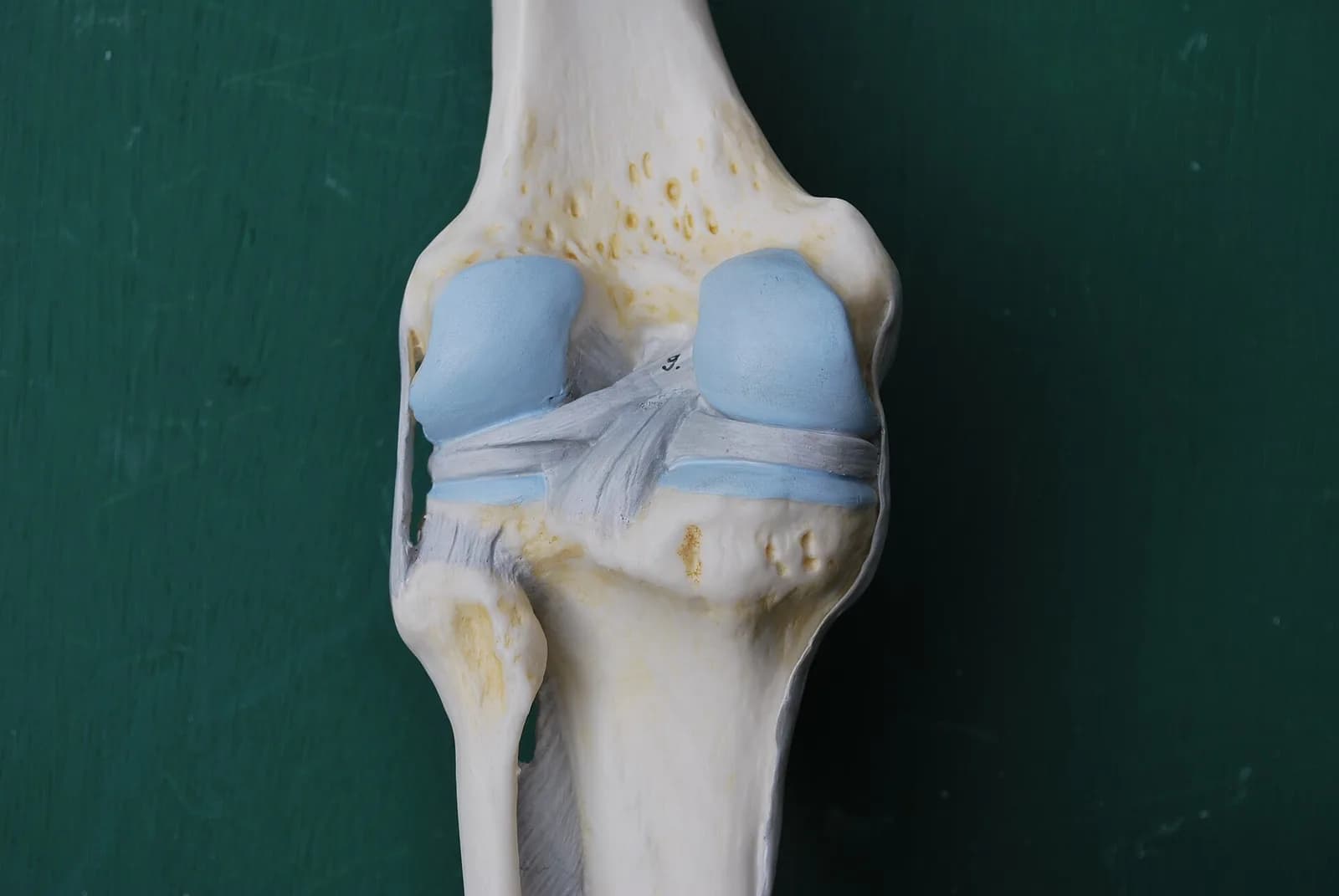
Cartilage in the body is not all the same1. Elastic cartilage is soft and flexible, while fibrocartilage is tougher and better at absorbing force1. The smooth, slippery tissue that allows bones to glide over one another is hyaline cartilage, also called articular cartilage, which is the type most commonly damaged by osteoarthritis1.
How did blocking 15-PGDH change cartilage cells?
Rather than using stem cells, the treatment prompted existing chondrocytes to alter which genes were active and take on a more youthful profile12.
In the single-cell analysis, one group of older chondrocytes that made 15-PGDH and showed gene activity for cartilage breakdown fell from 8% to 3%12. A second group that lacked 15-PGDH and expressed fibrocartilage-related genes declined from 16% to 8%1. Meanwhile, a third group that lacked 15-PGDH and expressed genes for forming hyaline cartilage increased from 22% to 42%1.
Single-cell analysis of mouse knee cartilage Unit: % of chondrocytes.
- 15-PGDH-producing degrading
- Fibrocartilage-forming
- Hyaline cartilage-forming
| Group | Bar | Value |
|---|---|---|
| Before treatment | 15-PGDH-producing degrading | 8 |
| Before treatment | Fibrocartilage-forming | 16 |
| Before treatment | Hyaline cartilage-forming | 22 |
| After treatment | 15-PGDH-producing degrading | 3 |
| After treatment | Fibrocartilage-forming | 8 |
| After treatment | Hyaline cartilage-forming | 42 |
The extracellular matrix consists of proteins and other molecules that surround cells and provide structural support1. Within cartilage, this matrix matters because it lets the tissue handle pressure and keeps the joint surface smooth for movement1.
What did the mouse experiments show?
After treatment, knee cartilage in older mice became thicker and consisted of the smooth articular cartilage that joints need to move without friction12. In the injury model, mice treated twice a week for four weeks after an ACL-type injury were less likely to develop osteoarthritis and put more weight on the injured leg than animals that did not receive the inhibitor1.
The published abstract reports that both systemic and local inhibition of 15-PGDH regenerated articular cartilage and reduced osteoarthritis-associated pain in mice2. By about 15 years after an ACL tear, roughly 50% of people have developed osteoarthritis in that joint1. Mice that received a control drug showed 15-PGDH levels twice as high as those of uninjured mice and developed osteoarthritis within four weeks1.
Researchers observed that cartilage regeneration to this extent in aged mice was surprising1. The treated mice also moved more normally than untreated animals1.
What happened in human cartilage?
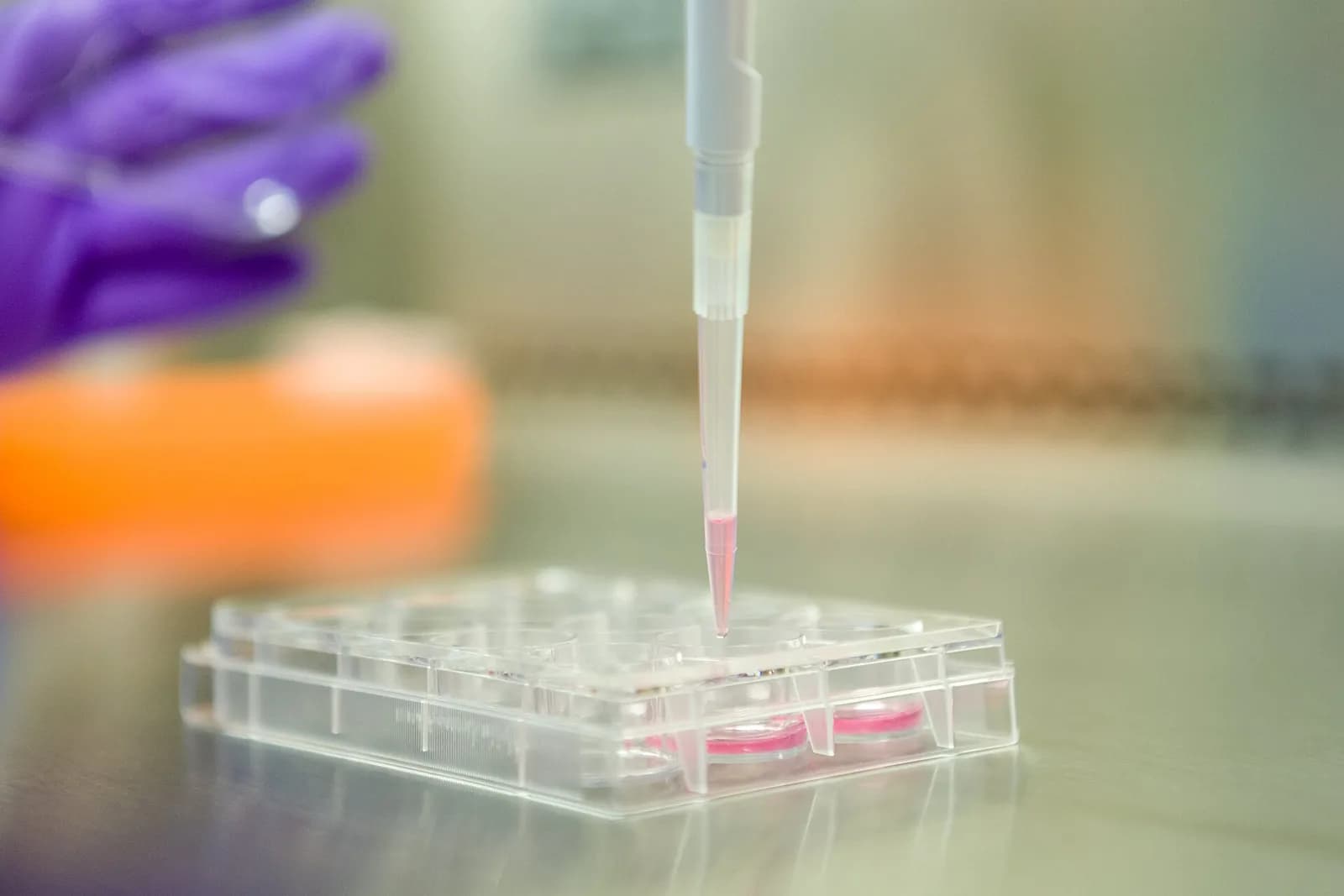
The group also tested cartilage removed from people with osteoarthritis during operations to replace the entire knee1. After one week of exposure to the inhibitor, treated samples contained fewer cells making 15-PGDH, and genes related to cartilage breakdown and fibrocartilage were also less active than in untreated tissue1.
The important observation was that the samples started to form new articular cartilage1. Even so, these results do not yet show that the approach regrows cartilage or prevents osteoarthritis in humans; researchers say clinical studies are required1. An oral 15-PGDH inhibitor has already reached early clinical testing for muscle weakness, and researchers hope a similar trial will be launched soon for cartilage regeneration1.
Frequently asked questions
What is 15-PGDH?
15-PGDH is an enzyme that breaks down prostaglandin E2 and becomes more abundant with age in mouse knee cartilage1. Stanford researchers describe it as a gerozyme, an enzyme linked to age-related tissue decline1.
Did the treatment work in humans?
Human cartilage samples from knee replacement surgeries responded to the inhibitor in the lab by producing new articular cartilage1. However, this was not a clinical trial; researchers say studies in people are still required1.